

利用淬灭荧光蛋白分子的化学再活化实现树脂嵌入式荧光显微成像
染色方法:
转基因荧光标记
标记方法:
GFP、 YFP、 RFP
包埋方法:
琼脂糖包埋
成像平台:
BioMapping 5000
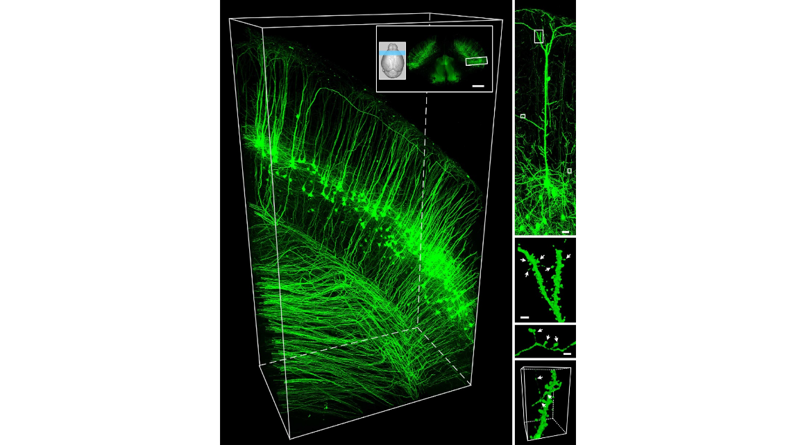
Figure 4 | Imaging of the thy1-YFPH mouse brain. (a) 3D presentation of a P21 thy1-YFPH mouse brain data set. Images were acquired with a voxel size of 0.5?0.5?1mm 3 . The inset image shows the contours of the whole mouse brain, reconstructed by autofluorescence and coronal plane sectioning on the blue labelled region (centred at the bregma þ1.50 level). Structures of the cerebral cortex are highlighted (white boxing inset) and enlarged to show details. The z axis represents the buffer penetration direction. (b) A layer Vpyramidal neuron recorded at a 0.2?0.2?0.2mm 3 voxel size on a commercial confocal microscope (Zeiss, LSM710). c–e show enlargements of the fine structures from the white box region of b. White arrows and numerals indicate (i) stubby, (ii) branched, (iii) thin, (iv) filopodium and (v) mushroom spines, as well as (vi) terminaux boutons, (vii) en passant bouton and (viii) structures suspected as synaptic connections. Scale bar: (a) inset, 1mm; (b), 20mm; (c,d), 2mm. Dimensions: (a) 1,850x620x1,000mm 3 ; (e) 9x18x9mm 3 .
Movie 1: Traveling in the brain
2014年6月2日,华中科技大学武汉光电国家研究中心骆清铭教授课题组发现绿色荧光蛋白在树脂包埋的过程中荧光重激活方法,使用fMOST技术,研究小组演示了如何获取密集的全脑神经网络数据。文章发表在《自然-通讯》杂志。
参考文献
参考文献[1]:Xiong H, Zhou Z, Zhu M, Lv X, Li A, Li S, Li L, Yang T, Wang S, Yang Z, Xu T, Luo Q, Gong H, Zeng S. Chemical reactivation of quenched fluorescent protein molecules enables resin-embedded fluorescence microimaging. Nat Commun. (2014);5:3992.