

塑性包埋大体积免疫标记样品的三维高分辨率成像
染色方法:
整体免疫荧光
标记方法:
AlexaFluor488 、CY2、 CY3、CY5、FITC
包埋方法:
树脂包埋
成像平台:
BioMapping 5000

cover
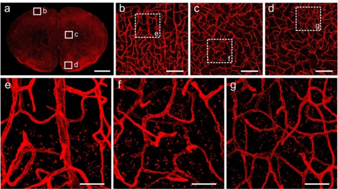
Fig. 4. Fluorescent images of Lectin-DyLight 594 labeled vasculature in the brain tissue after Lowicryl HM20 resin embedding. (a) Maximum intensity projections of a 20-μm-thick coronal slice. (b-d) Corresponding magnification of regions indicated in (a). (e-g) High magnification images of the boxed regions in (b-d), respectively. All images were acquired using 20 × (NA = 1.0), at a 0.42 × 0.42 × 1.00 μm 3 voxel size on a confocal microscope (LSM780, ZEISS). Scale bars: (a) 1 mm; (b-d) 100 μm; (e-g) 30 μm.
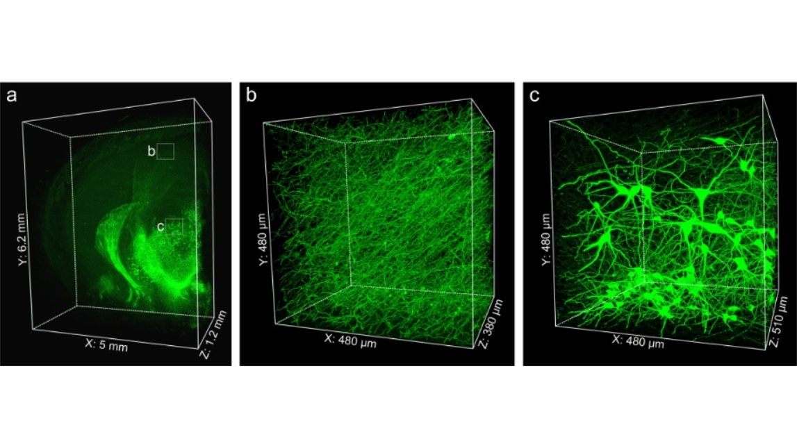
Fig. 5. Imaging large volume immunolabeled mouse brain tissue after Lowicryl HM20 resin embedding. (a) 3D presentation of the TH immunolabeled mouse brain block. Enlargements of the fine structures of TH-positive axonal fibers in the cortex (b) and TH-positive soma located in the thalamus (c). Images were acquired by successive high-resolution stage-scanning microscopy at a 0.16 × 0.16 × 1.00 μm 3 voxel size. (a) 6200 × 5000 × 1200 μm 3 ; (b) 480 × 480 × 380 μm 3 ; (c) 480 × 480 × 510 μm 3
cover
Fig. 4. Fluorescent images of Lectin-DyLight 594 labeled vasculature in the brain tissue after Lowicryl HM20 resin embedding. (a) Maximum intensity projections of a 20-μm-thick coronal slice. (b-d) Corresponding magnification of regions indicated in (a). (e-g) High magnification images of the boxed regions in (b-d), respectively. All images were acquired using 20 × (NA = 1.0), at a 0.42 × 0.42 × 1.00 μm 3 voxel size on a confocal microscope (LSM780, ZEISS). Scale bars: (a) 1 mm; (b-d) 100 μm; (e-g) 30 μm.
Fig. 5. Imaging large volume immunolabeled mouse brain tissue after Lowicryl HM20 resin embedding. (a) 3D presentation of the TH immunolabeled mouse brain block. Enlargements of the fine structures of TH-positive axonal fibers in the cortex (b) and TH-positive soma located in the thalamus (c). Images were acquired by successive high-resolution stage-scanning microscopy at a 0.16 × 0.16 × 1.00 μm 3 voxel size. (a) 6200 × 5000 × 1200 μm 3 ; (b) 480 × 480 × 380 μm 3 ; (c) 480 × 480 × 510 μm 3
2017年7月10日,华中科技大学武汉光电国家研究中心曾绍群教授课题组,提出了一种结合大样本树脂包埋和 iDISCO 免疫荧光染色的方法来获得高空间分辨率的生物分子的方法。 这一方法结合fMOST技术,能够以高分辨率成像免疫标记的大体积组织。文章发表在《Biomed Opt Express》杂志上。
参考文献
参考文献[1]:Gang Y, Liu X, Wang X, Zhang Q, Zhou H, Chen R, Liu L, Jia Y, Yin F, Rao G, Chen J, Zeng S. Plastic embedding immunolabeled large-volume samples for three-dimensional high-resolution imaging. Biomed Opt Express. (2017);8(8):3583-3596.