

小鼠皮层中单个神经元的稀疏标记以及全脑形态重建
染色方法:
病毒示踪标记
标记方法:
EGFP、EYFP、 ERFP、 mCherry、 Tdtomato
包埋方法:
树脂包埋
成像平台:
BioMapping 5000
cover
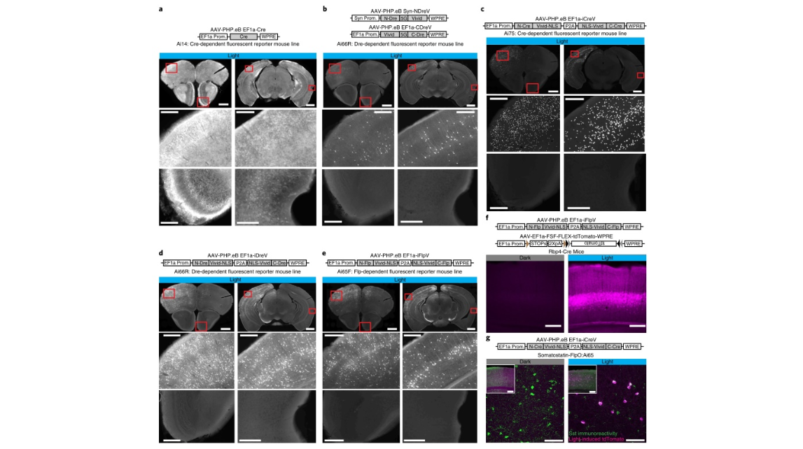
Fig. 2 | Optogenomic modifications with spatiotemporal and cell-class-specific precision in vivo. Reporter mice (n = 2 per case) received right hemisphere intracerebroventricular or retro-orbital injection of the indicated PHP.eB rAAVs followed by light stimulation to the left hemisphere 2 weeks postinjection, and imaging 2 weeks post-light stimulation. a, tdTomato expression in Ai14 mice injected with AAV-PHP.eB EF1a-Cre; 68,928 and 182,022 cells per section (CPS) were labeled. b, tdTomato expression in Ai66R mice ICV injected with a 1:1 mixture of AAV-PHP.eB Syn-NDreV and AAVPHP.eB EF1a-CDreV; 204 and 608 CPS were labeled. c, Nuclear-localized tdTomato expression in Ai75 mice ICV injected with AAV-PHP.eB EF1a-iCreV; 1,323 and 3,649 CPS were labeled. d, Ai66R mice were ICV injected with AAV-PHP.eB EF1a-iDreV (1,630 and 2,670 CPS). e, tdTomato reporter Ai65F mice were ICV injected with AAV-PHP.eB EF1a iFlpV. Scale bars (a–e), 1 mm for top images, 200 μm for bottom images (1,386 and 2,471 CPS). f, L5 pyramidal neuron-specific Rbp4-Cre mice were locally injected with a mixture of AAV-PHP.eB EF1a-iFlpV and AAV-PHP.eB EF1a-FSF-FLEX-tdTomato. Scale bars, 250 μm (469 of 478 cells in L5). g, Somatostatin FlpO mouse line, Sst-IRES-FlpO, crossed with a Cre/Flp double-dependent tdTomato reporter mouse line, Ai65, was retro-orbitally injected with AAV-PHP.eB EF1a-iCreV and light was delivered to the left hemisphere. Recombination was observed in somatostatin-positive inhibitory interneurons as revealed by immunohistochemistry (100% of reporter positive cells (119) were Sst positive (313); 38.1% of Sst cells were reporter positive). Scale bars, 250 μm for inset and 75 μm for zoomed images. All in vivo light activation was applied through the skull on the left hemisphere (opposite intracerebroventricular injection sites in those cases). For a–e, two coronal planes are shown for each injection (top row) with enlarged views (lower two rows) for areas indicated by red boxes. ICV, intracerebroventricularly
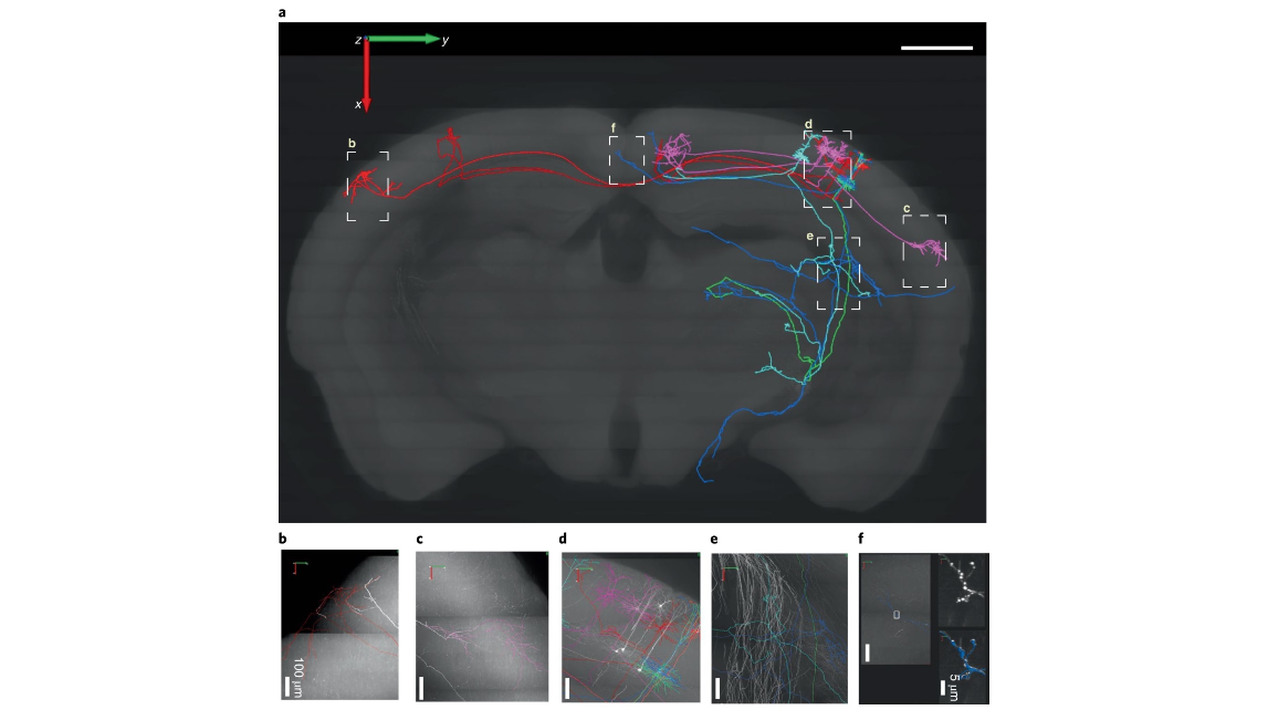
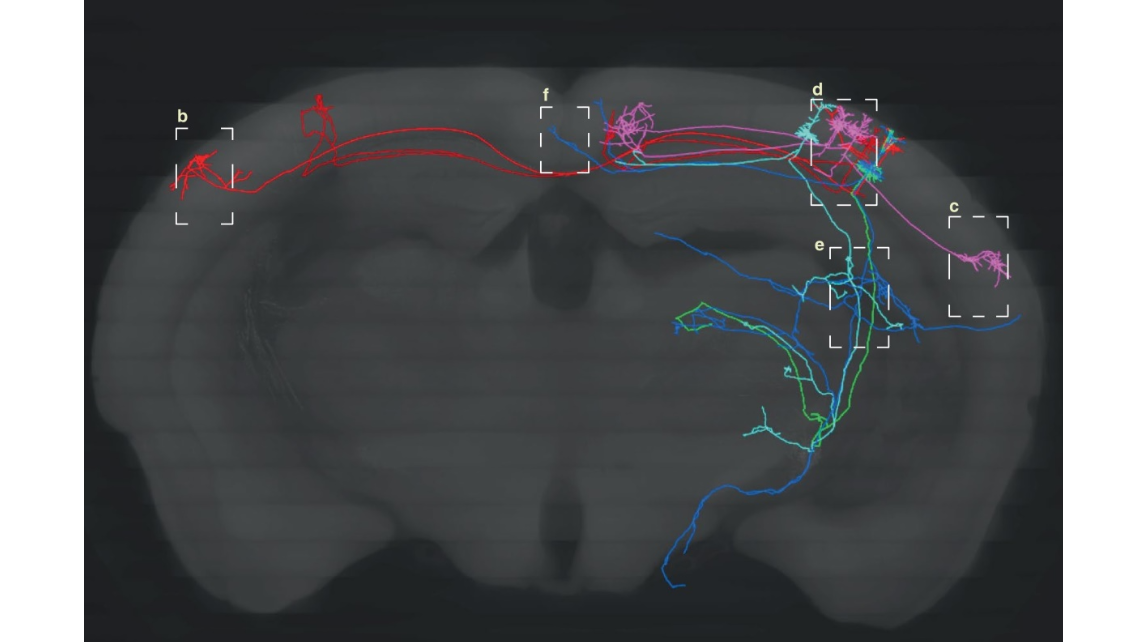
Fig. 4 | Cortical PCs labeled RecVs and were reconstructed at the whole-brain level. a, Eight reconstructed PCs in a mouse somatosensory cortex include three layer-2/3 PCs (in pink) with ipsilateral cortico-cortical projections, two layer-2/3 PCs (in red) with contralateral cortico-cortical projections and three L5 thick-tufted PCs (one green, one blue, one light blue) with ipsilateral cortico-subcortical projections. Local axonal clusters are incomplete because labeling at the somata region is too dense in this brain for tracing fine axonal branches. The eight reconstructed PCs are superimposed onto a coronal brain plane located 5,201–5,400 µm posterior to the olfactory bulb (scale bar, 1 mm). Five Ai139 EGFP reporter mice received local virus injection and light stimulation, and whole-brain neuronal reconstruction was performed using the fMOST images from one mouse that received 5 min of light stimulation. b–f, Enlarged views of areas outlined by dashed boxes in a, with reconstructions (in colors) superimposed on original images with EGFP fluorescence shown as white. In f, the two panels on the right (without reconstruction in white, with reconstruction in blue) are enlarged views of the boxed area in the left panel, showing the high-resolution details of a segment of axon with enlarged boutons. The whole-brain image stack is composed of 12,089 images; resolution of xyz, 0.3 × 0.3 × 1 µm3.
mov 1. Mouse visual cortical PCs reconstructed at whole-brain level; horizontal axis rotation. Eight EGFP-labeled neurons were imaged by fMOST and reconstructed from barrel cortex of a mouse brain, and are presented in 3D within a partial brain contour composed of serial reconstructed contours of coronal brain sections. The eight PCs include three L2/3 PCs (in pink) having ipsilateral cortico-cortical projections, two L2/3 PCs (in red) having contralateral cortico-cortical projections and three L5 TTPCs (thick-tufted PCs, one in green, one in blue, one in light blue) having cortico-subcortical projections. Note: local axonal clusters are incomplete because the labeling at the region around their somata is too dense for tracing fine axonal branches.
mov 2. Mouse visual cortical PCs reconstructed at whole-brain level; vertical axis rotation. Eight EGFP-labeled neurons were imaged by fMOST and reconstructed from barrel cortex of a mouse brain, and are presented in 3D within a partial brain contour composed of serial reconstructed contours of coronal brain sections. The eight PCs include three L2/3 PCs (in pink) having ipsilateral cortico-cortical projections, two L2/3 PCs (in red) having contralateral cortico-cortical projections and three L5 TTPCs (thick-tufted PCs, one in green, one in blue, one in light blue) having cortico-subcortical projections. Note: local axonal clusters are incomplete because the labeling at the region around their somata is too dense for tracing fine axonal branches. Supplementary Information
cover
Fig. 2 | Optogenomic modifications with spatiotemporal and cell-class-specific precision in vivo. Reporter mice (n = 2 per case) received right hemisphere intracerebroventricular or retro-orbital injection of the indicated PHP.eB rAAVs followed by light stimulation to the left hemisphere 2 weeks postinjection, and imaging 2 weeks post-light stimulation. a, tdTomato expression in Ai14 mice injected with AAV-PHP.eB EF1a-Cre; 68,928 and 182,022 cells per section (CPS) were labeled. b, tdTomato expression in Ai66R mice ICV injected with a 1:1 mixture of AAV-PHP.eB Syn-NDreV and AAVPHP.eB EF1a-CDreV; 204 and 608 CPS were labeled. c, Nuclear-localized tdTomato expression in Ai75 mice ICV injected with AAV-PHP.eB EF1a-iCreV; 1,323 and 3,649 CPS were labeled. d, Ai66R mice were ICV injected with AAV-PHP.eB EF1a-iDreV (1,630 and 2,670 CPS). e, tdTomato reporter Ai65F mice were ICV injected with AAV-PHP.eB EF1a iFlpV. Scale bars (a–e), 1 mm for top images, 200 μm for bottom images (1,386 and 2,471 CPS). f, L5 pyramidal neuron-specific Rbp4-Cre mice were locally injected with a mixture of AAV-PHP.eB EF1a-iFlpV and AAV-PHP.eB EF1a-FSF-FLEX-tdTomato. Scale bars, 250 μm (469 of 478 cells in L5). g, Somatostatin FlpO mouse line, Sst-IRES-FlpO, crossed with a Cre/Flp double-dependent tdTomato reporter mouse line, Ai65, was retro-orbitally injected with AAV-PHP.eB EF1a-iCreV and light was delivered to the left hemisphere. Recombination was observed in somatostatin-positive inhibitory interneurons as revealed by immunohistochemistry (100% of reporter positive cells (119) were Sst positive (313); 38.1% of Sst cells were reporter positive). Scale bars, 250 μm for inset and 75 μm for zoomed images. All in vivo light activation was applied through the skull on the left hemisphere (opposite intracerebroventricular injection sites in those cases). For a–e, two coronal planes are shown for each injection (top row) with enlarged views (lower two rows) for areas indicated by red boxes. ICV, intracerebroventricularly
Fig. 4 | Cortical PCs labeled RecVs and were reconstructed at the whole-brain level. a, Eight reconstructed PCs in a mouse somatosensory cortex include three layer-2/3 PCs (in pink) with ipsilateral cortico-cortical projections, two layer-2/3 PCs (in red) with contralateral cortico-cortical projections and three L5 thick-tufted PCs (one green, one blue, one light blue) with ipsilateral cortico-subcortical projections. Local axonal clusters are incomplete because labeling at the somata region is too dense in this brain for tracing fine axonal branches. The eight reconstructed PCs are superimposed onto a coronal brain plane located 5,201–5,400 µm posterior to the olfactory bulb (scale bar, 1 mm). Five Ai139 EGFP reporter mice received local virus injection and light stimulation, and whole-brain neuronal reconstruction was performed using the fMOST images from one mouse that received 5 min of light stimulation. b–f, Enlarged views of areas outlined by dashed boxes in a, with reconstructions (in colors) superimposed on original images with EGFP fluorescence shown as white. In f, the two panels on the right (without reconstruction in white, with reconstruction in blue) are enlarged views of the boxed area in the left panel, showing the high-resolution details of a segment of axon with enlarged boutons. The whole-brain image stack is composed of 12,089 images; resolution of xyz, 0.3 × 0.3 × 1 µm3.
mov 1. Mouse visual cortical PCs reconstructed at whole-brain level; horizontal axis rotation. Eight EGFP-labeled neurons were imaged by fMOST and reconstructed from barrel cortex of a mouse brain, and are presented in 3D within a partial brain contour composed of serial reconstructed contours of coronal brain sections. The eight PCs include three L2/3 PCs (in pink) having ipsilateral cortico-cortical projections, two L2/3 PCs (in red) having contralateral cortico-cortical projections and three L5 TTPCs (thick-tufted PCs, one in green, one in blue, one in light blue) having cortico-subcortical projections. Note: local axonal clusters are incomplete because the labeling at the region around their somata is too dense for tracing fine axonal branches.
mov 2. Mouse visual cortical PCs reconstructed at whole-brain level; vertical axis rotation. Eight EGFP-labeled neurons were imaged by fMOST and reconstructed from barrel cortex of a mouse brain, and are presented in 3D within a partial brain contour composed of serial reconstructed contours of coronal brain sections. The eight PCs include three L2/3 PCs (in pink) having ipsilateral cortico-cortical projections, two L2/3 PCs (in red) having contralateral cortico-cortical projections and three L5 TTPCs (thick-tufted PCs, one in green, one in blue, one in light blue) having cortico-subcortical projections. Note: local axonal clusters are incomplete because the labeling at the region around their somata is too dense for tracing fine axonal branches. Supplementary Information
2020年3月23日,美国艾伦脑科学研究所、斯坦福大学联合中国华中科技大学、温州医科大学等,利用RecV重组酶系统实现了对单细胞或细胞群体的体内靶向光诱导基因组修饰。本研究中,与fMOST技术结合,使得小鼠皮层中单个神经元的稀疏标记以及全脑形态重建成为可能。文章发表在《自然-方法》杂志。
参考文献
参考文献[1]:Shenqin Yao et al., RecV Recombinase System for in Vivo Targeted Optogenomic Modifications of Single Cells or Cell Populations. Nat Methods 2020 Mar 23 [Online ahead of print]