

三维分析脑内出血小鼠神经元凋亡和血管生成
染色方法:
尼氏染色
标记方法:
Nissl染色
包埋方法:
树脂包埋
成像平台:
BioMapping 1000
cover

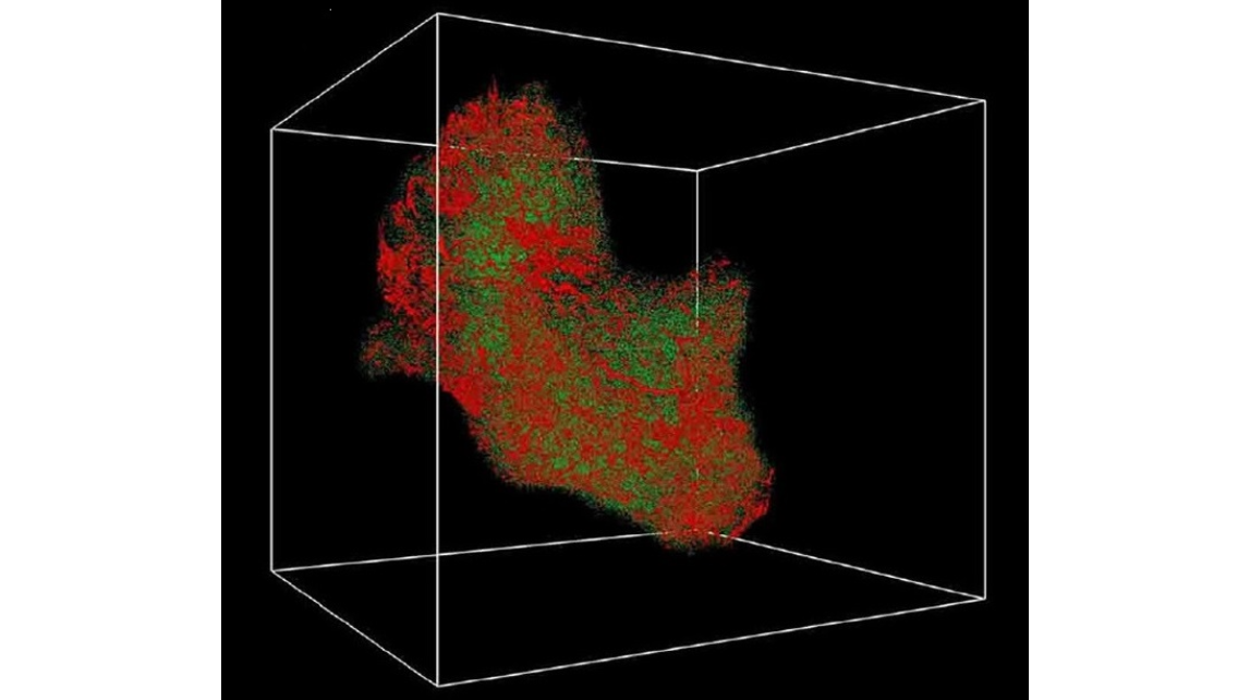
figure 2. Representative 3D reconstruction video screenshot of a mouse brain in the vehicle and EGb761 groups, respectively.The regions in the video covered the lesion area (b, d), and the contralateral area (a, c). From the screenshot images, we can find that the microvessel density was higher in the lesions of the EGb761-treated mice, when compared with the vehicle group.
video 1
video 2
video 3
video 4
cover
figure 2. Representative 3D reconstruction video screenshot of a mouse brain in the vehicle and EGb761 groups, respectively.The regions in the video covered the lesion area (b, d), and the contralateral area (a, c). From the screenshot images, we can find that the microvessel density was higher in the lesions of the EGb761-treated mice, when compared with the vehicle group.
video 1
video 2
video 3
video 4
2018年2月5日,华中科技大学同济医学院同济医院神经内科唐洲平教授课题组,采用MOST获取脑出血小鼠神经血管网络的图像数据集,揭示银杏叶提取物 EGb761对实验性脑出血神经元凋亡的保护作用。文章发表在《分子神经生物学》杂志上。
参考文献
参考文献[1]:Pan C, Liu N, Zhang P, Wu Q, Deng H, Xu F, Lian L, Liang Q, Hu Y, Zhu S, Tang Z. EGb761 Ameliorates Neuronal Apoptosis and Promotes Angiogenesis in Experimental Intracerebral Hemorrhage via RSK1/GSK3β Pathway. Mol Neurobiol. (2017). doi: 10.1007/s12035-016-0363-8.